Software to Automatically Detect Ellipsoid Zone Loss in SD-OCT Imaging
Ellipsoid zone (EZ) loss has been proposed as evidence of progression of several retinal degenerative diseases, including, but not limited to, retinitis pigmentosa and hydroxychloroquine (HCQ)-induced retinal toxicity. HCQ is a first-line drug used to treat autoimmune diseases such as systemic lupus erythematosus, Sjögren’s syndrome, and rheumatoid arthritis. HCQ-induced retinal toxicity is estimated to occur in 7.5% of patients taking the drug for more than 10 years, however, there is currently no treatment available.
NIH inventors have developed a method to automatically detect and quantify EZ loss in Spectral Domain-Optical Coherence Tomography (SD-OCT) images within seconds after image acquisition. This method includes a deep learning framework with a two-step approach.
In the first stage, the method detects and annotates EZ loss regions in individual OCT B-scans. A 2D map is constructed twice in a dual architecture to enhance robustness, where horizontal and vertical slices extracted from the 3D image are trained separately.
The second stage of the model operates on these two 2D maps and approximates the final EZ loss map representing the 3D OCT volume. 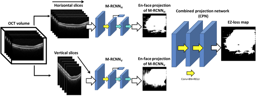
Compared to other screening methods, the algorithm demonstrated excellent performance in diagnosing toxicity even as a stand-alone test, with an F1 score, a measure of test accuracy, of 0.91. This indicates the utility of the tool in assisting with screening for toxicity in an automatic, accurate, time-effective, cost-effective, and objective manner. The addition of this methodology onto current SD-OCT screening could assist the clinician in making diagnostic and treatment decisions immediately after SD-OCT acquisitions.
Potential commercial applications of this software include:
- Methodology to integrate with SD-OCT imaging for screening applications for different retinal degeneration diseases.
- Implementation of this automatic algorithm also in screening outside of ophthalmology offices (OCTs become more ubiquitous in internal medicine settings).
- Quantitative data produced from the algorithms could provide surrogate endpoints for use in clinical trials and interventional studies aimed at halting the progression of degenerative changes.
For further information or to contact the licensing manager, please view the abstract: Systems and Methods to Automatically Detect Ellipsoid Zone Loss in SD-OCT Imaging.