
Quantitative Particle Identification (QPID) Digital Autoradiography System
Summary:
The National Cancer Institute (NCI) seeks research co-development partners and/or licensees for a Quantitative Particle Identification (QPID) digital autoradiography system with particle identification capabilities.
Description of Technology:
Autoradiography is a photographic process that exposes a medium, sensitive to radiation, to a sample emitting radiation. The current state of the art involves exposing a thin slice of tissue to an ionization-sensitive film for several hours or days and then scanning the film into an autoradiography image. This image is created using the sum of the ionizations from all the decays that occurred during the time of exposure on the ionization-sensitive medium. Imaging quality of these samples can be improved by collecting information from each individual decay to enhance the autoradiographic measurement.
Researchers at National Cancer Institute (NCI) developed a Quantitative Particle Identification (QPID) digital autoradiography system to measure the energy deposition from charged particles for each individual radioactive decay. The QPID leverages the ionizing radiation detection features of the Timepix3 detector to generate autoradiograph images in units of dose per unit time and area. The high readout speed of the Timepix3 gives the detector the capability to measure separate decay ionizations. It also segregates the image into one generated only by alpha and the other only by beta particles. A gamma detector was interfaced with the Timepix3 – allowing tagging charged particle ionization events in coincidence with a gamma emission during the isotope decay. This allows for a method to distinguish between alpha and positron emitting radioisotopes when imaged concurrently in the same pathology sample. This is the most unique feature of the QPID which will aid the development of new theranostic treatments in which two similar ligands are used.
Researchers at the NCI seek licensing and/or co-development research collaborations to continue developing the QPID system for improved autoradiography imaging.
Potential Commercial Applications:
- Improved radiotherapy dosing for cancer treatment
- Improved radiotherapy guidance for cancer treatment
- Improve accuracy of biopsy procedures through real-time tracer measurement and detection
- Measuring dual radioligand pathology samples
Competitive Advantages:
- QPID can measure charge particle activity for individual decays (alpha, beta, and gamma)
- QPID measures absolute time of decay relative to the start of data acquisition
- QPID more accurately measures radioactive dose in a tissue sample
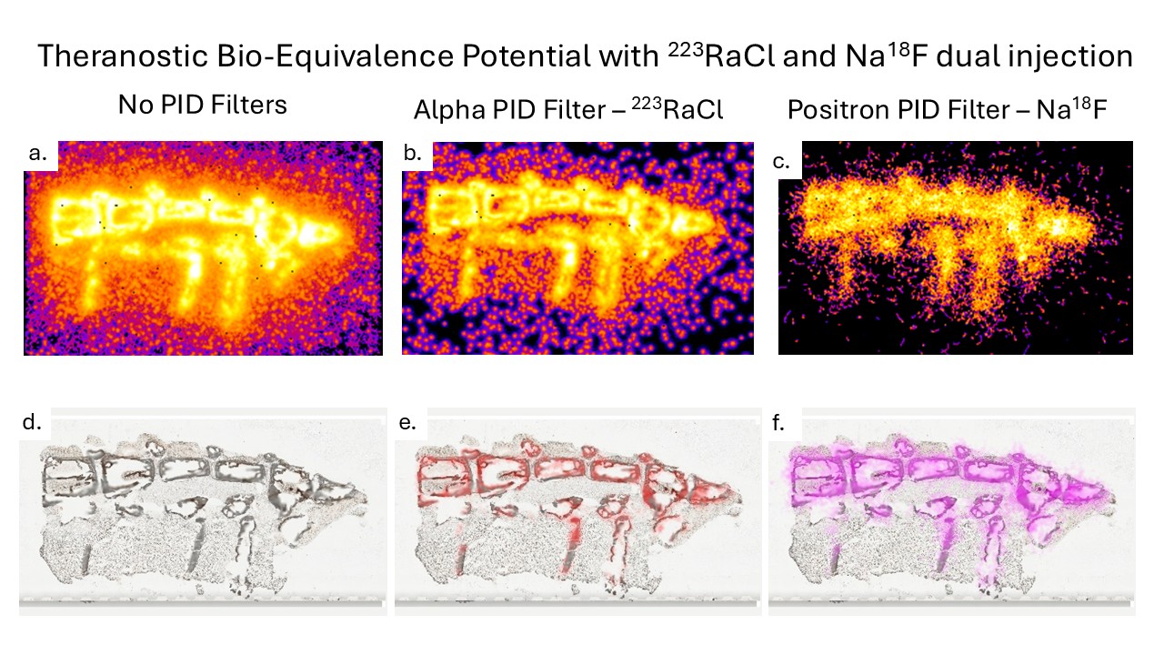
The figure shows the unique ability of the QPID to image two radioisotopes simultaneously. A mouse was injected with a mixture of 223RaCl and Na18F, a theranostic agent pair which treats and images bone metastasis from prostate cancer. An hour after injection, a 50 micron thick spine sample was imaged on the QPID. The 223RaCl and Na18F uptake into the bone was separated into two images, one as the distribution of 223RaCl and the other as the distribution of Na18F. Because the QPID records individual decays of either radioisotope, accurate amounts of the two radioisotopes can be measured and studies of the biodistribution and micro-dosimetry are possible. The top row (panels a, b, and c) are autoradiography images generated by the QPID. The bottom row (panels d, e and f) are co-registered images of photographs of the sample overlayed with the radioisotope distribution.